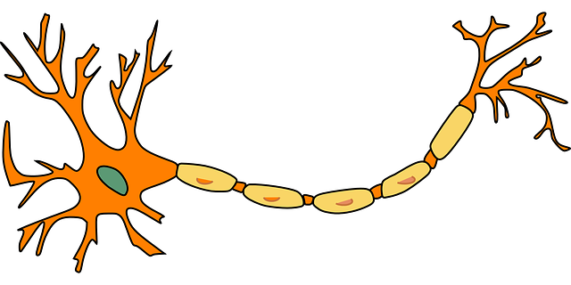
How do scientists study the activity of cells in the brain? If light could not reach the skull, how would experimenters see neurons in the brain? There are billions of neurons. How do investigators tell which neurons are active while monitoring many neurons at once? Before looking into the questions above, let’s walk through a scenario together!
Imagine that you are in a forest and want to know whether a precious animal lives in it. What would you do? An experienced biologist may start by searching for the traces that the animal could have left – such as footprints, scratches on the trees, or feces. The existence of those signs would tell the biologist that there is a great chance of finding that animal in the forest. Neuroscientists have adopted a similar strategy to identify the occurrence of neural activity.
Then, what traces does a neuron leave when it turns on? Imagine that each neuron is a castle. When the neuron is in the “off mode”, the door of the castle is locked. Potassium ions, which are the little princes and princesses, are required to stay inside the castle. Sodium ions, the commoners who live outside the castle, are banned from entering. This segregation of nobles and commoners makes both sides eager to explore each other’s world.
The good news is that the castle does not always stay closed. Whenever important information from other kings arrives at the castle, the neuron will switch to the “on mode” and open its door. During the open house event, potassium ions will leave the castle to meet their commoner friends, and the sodium ions will enter the castle to experience the nobles’ luxurious lives. At the same time, calcium ions, which are the wise people among commoners, will be invited to the castle and will offer their advice to the king.
Based on the analogy above, what traces could you use to tell whether a castle is holding an open house event or not? Scientists selected two traces:
-
the entering of the calcium ions (i.e., wise people) into the neuron
-
the increase in voltage across the cell membrane caused by the movement of potassium and sodium ions
Scientists “see” calcium ions using GCaMP, a type of calcium indicator (Chen et al., 2012; Chen et al., 2013). It lights up with fluorescent signals when it detects calcium ions in the neuron. Because calcium ions generally only enter a neuron when it is in the “on mode”, seeing a neuron light up tells researchers that the neuron is activated by some signal at that time. In the video below, a group of investigators used GCaMP to record the activity of a structure called the hippocampus in a mice’s brain while the mice was participating in an experiment (Modi et al., 2014). Notice that some neurons were flashing like stars – they are helping the mice complete a behavioral task!
QuasAr is a type of voltage indicator (Hochbaum et al., 2014). The strength of the fluorescent signals it emits depends on the membrane voltage – the higher the voltage, the brighter the indicator! In the video below, the researchers artificially activated the neuron in the frame (Hochbaum et al., 2014). You can see the change in the membrane voltage throughout the neuron as it switches on and quickly turns off. Isn’t it just like a beautiful firework?
Notice in the paragraph above, I said “the researchers artificially activated the neuron.” As you may have guessed, the investigators achieved that by controlling the gate of the castle. Channelrhodopsin is a gate that could be opened by blue light (Nagel et al., 2003). It can be artificially installed on castles. The opening of this gate allows the ions to pass through, forcing the neuron to switch to the “on mode”. Go back to the video above and find the blue-shaded area – when the blue light was shined on this part of the neuron, it turned on!
Similar to the mechanism of channelrhodopsin, halorhodopsin is an artificial gate that could be opened by yellow or red light (Gradinaru et al., 2010). Only chloride ions, which are the evil assassins belonging to the other kings, could go through the gate. Thus, opening this gate will increase the castle’s alertness. This makes it harder to switch a neuron into the “on mode” because the castle does not want to open its main gate unless the information it is receiving is super important. The two types of gates, channelrhodopsin and halorhodopsin, can be installed on the same neuron. This allows researchers to turn the neuron on or off as they wish (Gradinaru et al., 2010)!
credit: https://giphy.com/123pingu/
I hope you enjoyed reading this article and learning about cool neuroscience technologies. What I have covered here is just a small portion of all of the technologies available. But I believe that they serve as a great demonstration of the beauty (both conceptually and literally) of neuroscience research!
References
Chen Q, Cichon J, Wang W, Qiu L, Lee SJ, Campbell NR, Destefino N, Goard MJ, Fu Z, Yasuda R, Looger LL, Arenkiel BR, Gan WB, Feng G. (2012). Imaging neural activity using Thy1-GCaMP transgenic mice. Neuron, 76(2):297-308. doi: 10.1016/j.neuron.2012.07.011.
Chen TW, Wardill TJ, Sun Y, Pulver SR, Renninger SL, Baohan A, Schreiter ER, Kerr RA, Orger MB, Jayaraman V, Looger LL, Svoboda K, Kim DS. (2013). Ultrasensitive fluorescent proteins for imaging neuronal activity. Nature, 499(7458):295-300. doi: 10.1038/nature12354.
Gradinaru, V., Zhang, F., Ramakrishnan, C., Mattis, J., Prakash, R., Diester, I., Goshen, I., Thompson, K. R., & Deisseroth, K. (2010). Molecular and cellular approaches for diversifying and extending optogenetics. Cell, 141(1):154–165. doi: 10.1016/j.cell.2010.02.037.
Hochbaum, D. R., Zhao, Y., Farhi, S. L., Klapoetke, N., Werley, C. A., Kapoor, V., Zou, P., Kralj, J. M., Maclaurin, D., Smedemark-Margulies, N., Saulnier, J. L., Boulting, G. L., Straub, C., Cho, Y. K., Melkonian, M., Wong, G. K., Harrison, D. J., Murthy, V. N., Sabatini, B. L., Boyden, E. S., … Cohen, A. E. (2014). All-optical electrophysiology in mammalian neurons using engineered microbial rhodopsins. Nature methods, 11(8): 825–833. doi: 10.1038/nmeth.3000.
Modi, M. N., Dhawale, A. K., & Bhalla, U. S. (2014). CA1 cell activity sequences emerge after reorganization of network correlation structure during associative learning. eLife, 3(e01982). doi: 10.7554/eLife.01982.
Nagel, G., Szellas, T., Huhn, W., Kateriya, S., Adeishvili, N., Berthold, P., Ollig, D., Hegemann, P., & Bamberg, E. (2003). Channelrhodopsin-2, a directly light-gated cation-selective membrane channel. PNAS, 100(24):13940-13945. doi: 10.1073/pnas.1936192100.